
ALINA M. PICOS,1 SIMINA POENAR,1 ALEXANDRA OPRIS,2 ALEXANDRA CHIRA,2 MARIUS BUD,3ANTONELA BERAR,1 ANDREI PICOS,1 and DAN L. DUMITRASCU2
Abstract:
Background
Dental erosions are determined by a mechanism involving increased oral acidity. Gastro-esophageal reflux disease (GERD) represents the pathological reflux of gastric content into the oral cavity, affecting the hard dental tissues integrity, with a major risk of advanced tooth wear.
Aim
This study aims to investigate the prevalence of dental erosions in GERD patients, in order to obtain a basis for therapeutic strategies and specific prophylactic measures.
Methods
We incorporated a mandatory dental consultation in the therapeutic protocol of GERD patients. The study was carried out in a group of 60 patients with GERD. Dental examination of these patients revealed 21 cases showing visible dental erosions. The control group included 60 patients, without GERD, age and sex matched. All examinations were carried out in a tertiary center by the same team of dentists, instructed in dental erosion recognition and questionnaire administration.
Results
The dental erosion prevalence in patients with GERD was 35%. In the control group, erosions were 13% (OR: 3.6); 67% of patients with dental erosions were females and 33% were males. Middle age showed the highest risk for dental erosions, with peaks in the fourth and sixth decades.
Conclusions
The risk of dental erosion was significantly higher in GERD patients as compared to the control group. The sex ratio shows a higher prevalence of erosion in females
Keywords: dental erosion, GERD
Introduction
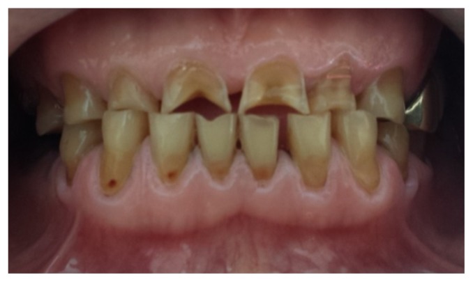
The erosion of tooth structure is defined as a loss of dental hard tissue surface due to a chemical process of acid attack that does not involve bacterial factors (Fig 1). Exogenous factors are involved, related to the consumption of acidic foods and drinks, but also internal factors that decrease the pH in the oral cavity, which are common in gastro-esophageal pathology and hyperemesis [1].
Gastro-esophageal reflux disease (GERD) is a relatively common disorder, affecting daily approximately 7% of the adult population and 36% at least once a month [2]. GERD is caused by several mechanisms, mainly the abnormal pressure of the lower esophageal sphincter, allowing the gastric fluid to enter into the esophagus and even to reach the cervical portion of the esophagus, the pharynx or the oral cavity [2,3]. When a substance having a lower pH than 5.5, which is the critical pH for the enamel integrity, reaches the oral cavity, the crystals in the enamel hydroxyapatite can be dissolved. Gastric secretion has a pH between 0.8–2, with a great erosive potential [4].
Frequent exposure of teeth to this acid can cause severe tooth wear (erosion). Dentists are often the first healthcare providers for guiding the diagnosis of GERD due to the identification of dental erosion that might be the first sign of disease [5,6].
The loss of tooth hard structure leads to: hypersensitivity to thermal, tactile or sweet stimuli, pain during chewing, loss of the vertical dimension of occlusion with esthetic consequences due to the decrease of the lower 1/3 of the face [7].However, dental erosion can be a multifactorial phenomenon in which the protective buffering capacity of the saliva is exceeded by either a reduced salivary secretion, or a larger amount of regurgitated gastric fluid [8,9]. Normaly, in one to two hours after the acid attack, the remineralization is realized due to salivary calcium and phosphate ions [7].
Aim of the study
We started from the idea that oral acidity triggers erosion mechanisms; our purpose was to evaluate the severity of dental erosion risk in patients with GERD compared with healthy controls. Therefore we incorporated a mandatory dental consultation in the therapeutic protocol of GERD.
Materials and Methods
All patients with GERD symptoms referred to a tertiary center (3rd Medical Unit of the 2nd Medical Dept., Iuliu-Hatieganu University of Medicine and Pharmacy, Cluj-Napoca) were selected for the study.
After informed consent, all patients were submitted to clinical investigation, including a validated questionnaire on symptoms, upper digestive endoscopy, 24 hours pH monitoring and dental examination.
All examinations were performed in the university locations by the same team of dentists instructed in erosion recognition and questionnaire administration. The investigation of salivary qualities concerning acidity level, buffering capacity and flux debit, was made using specific salivary tests.
Photos were taken of patients showing dental erosions in order to observe the evolution process.
The study was carried out in a group of 60 patients during December 2012–December 2013.
A control group involved 60 patients, without GERD, matched for age and sex.
Each patient completed a specific questionnaire, which included two parts: the first part aimed at identifying and characterizing dental erosion using the BEWE index, and the second part investigated the symptoms of gastroesophageal reflux.
We used the BEWE index (Basic Erosion Wear Examination) for clinical evaluation, an internationally accepted index classification of dental erosion with the advantage of a simple system that can assess the severity of dental erosion without allowing different interpretations between evaluators.
Tooth examined surfaces were classified according to four levels of erosion severity:
0-no tooth wear
1-superficial loss of enamel
2-loss of less than 50% of the dental surface with frequently dentin exposure
3-dental tissues loss is more than 50% of the dental surface with frequent dentin exposure
The final score was obtained by summing the values o f the six examined sextants. According to the value of the final BEWE score, the patient was assigned a certain level of risk. The results of BEWE index provides a practical guide to the therapeutic management of dental erosions [4].
Patients with dentures were not included in the study, because we could not evaluate the BEWE score.
The investigation protocol and questionnaires were approved by local ethical committee.
Results and discussion
Dental examination of GERD patients revealed 21 cases presenting visible dental erosions. Thus, the dental erosion prevalence in patients with gastro-esophageal reflux was 35% of all patients with GERD, as shown in fig. 2.

The prevalence of dental erosion in the control group was 13% (n=8), significantly lower than in the GERD group (OR: 3.6).
Sex distribution of patients with gastro-esophageal reflux and erosions shows a sex ratio of 2:1; we detected erosions in 14 females and 7 males (see fig. 3).

The sex ratio shows a higher prevalence of erosion in females, in the group of 60 GERD patients, even if the prevalence of gastro-esophageal reflux disease is higher in males
Regarding age distribution we found a greater prevalence of dental erosions in the groups of age 41–50 years and 61–70 years (Fig 4). In the elderly, total dentures are very common, and we did not consider these patients for the analysis.

Approximately similar results of dental erosion in the control group were found by Muñoz 12.5% but he found erosions in 47.5% of the GERD group of patients [9].
Studies carried out in others geographic locations show significant differences: Oginni’s study in a Nigerian population found significant lower values of 16% dental erosion prevalence in GERD patients and 5% in controls [10].
Regarding GERD incidence, Schroeder found GERD in 15 patients of 20 [11] compared to Jones who found 60% in European population [12].
This is to our knowledge the first study in this area oriented to the investigation of dental erosions in GERD. We were able to report in this pilot study a high prevalence of dental erosions in GERD, which was higher in females than in males and in the middle aged patients.
These results emphasize the necessity to extensively evaluate patients with GERD and the dental check-up should never been omitted.
Dentists are often the first healthcare providers for guiding the diagnosis of GERD due to the identification of dental erosions that might be the first sign of disease. Early stages of erosion usually remain undiagnosed, therefore the establishment of early treatment may be missed.
Conclusions
In this study dental erosions were encountered in 35% of patients with GERD. The risk of dental erosion was significantly higher, than in an age and sex matched control patients (OR:3.6). Concerning the sex ratio, dental erosions were more frequent in females with GERD than in males (2:1). The age groups more likely to present dental erosions were the group of 41–50 years and the group of 61–70 years old.
Acknowledgment
This study is supported by an international AUF project coordinated by Iuliu Hatieganu University of Medicine and Pharmacy, Cluj-Napoca, Romania, during 2012–2014.
References
1. Fairburn CG, Stein A, Jones R. Eating habits and eating disorders during pregnancy. Psychosom Med. 1992;54(6):665–672. [PubMed]
2. Monnikes H, Bardhan KD, Stanghellini V, Berghöfer P, Bethke TD, Armstrong D. Evaluation of GERD symptoms during therapy. Digestion. 2007;75(Suppl 1):41–47. [PubMed]
3. Touyz SW, Liew VP, Tseng P, Frisken K, Williams H, Beumont PJ. Oral and dental complications in dieting disorders. J Eating Disorders. 1993;14(3):341–347. [PubMed]
4. Lussi A, Jaeggi T. Quintessence International, editor. Dental erosion Diagnosis, risk assessment, prevention, treatment. Paris: 2012.
5. Bartlett D. Intrinsic cause of erosion. Monograph Oral Sci Basel Karger. 2006;20:119–139. [PubMed]
6. Gilmour AG, Beckett HA. The voluntary reflux phenomenon. Br Dent J. 1993;175(10):368–372. [PubMed]
7. Touyz LZ, Stern J. Hypersensitive dentinal pain Attenuation with Potassium Nitrate. General Dent. 1999;47(1):42–45. [PubMed]
8. Touyz LZ. The acidity (pH) and buffering capacity of Canadian fruit juice and dental implications. J Can Dent Assoc. 1994;60(5):454–458. [PubMed]
9. Muñoz JV, Herreros B, Sanchiz V, et al. Dental and periodontal lesions in patients with gastro-oesophageal reflux disease. Dig Liver Dis. 2003;35(7):461–467. [PubMed]
10. Oginni AO, Agbakwuru EA, Ndububa DA. The prevalence of dental erosion in Nigerian patients with gastro-oesophageal reflux disease. BMC Oral Health. 2005;5(1):123–129. [PMC free article] [PubMed]
11. Schroeder PL, Filler SJ, Ramirez B, Lazarchik DA, Vaezi MF, Richter JE. Dental erosion and acid reflux disease. Ann Intern Med. 1995;122(11):809–815. [PubMed]
12. Jones R, Lydeard S. Prevalence of symptoms and dyspepsia in the community. Br Med J. 1989;298:30–32. [PMC free article] [PubMed]
Articles from Clujul Medical are provided here courtesy of Universty of Medicine and Pharmacy of Cluj-Napoca, Romania